| Parts of the Breast
Skin and capsules
Although we have covered the various protective covers in the sections above it is worth while reviewing the coverings of the breast with a relative perspective in relation to each other.
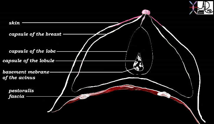
Each unit of the breast has a covering. The skin represents the first cover. The chest wall with the pectoralis muscle and its fascia represent the outer posterior cover of the breast. The capsule of mammary apparatus is the second cover, while the capsules of the individual lobes represent the third cover. Each lobule has a capsule and each acinus has a basement membrane which acts as a barrier or “capsule”. Each cell of course has a cell membrane.
| Parts of the breast |
|
This diagram reveals each cover of the progressively smaller units of the breast, including the skin, and capsules of the mammary apparatus, lobe, lobules, and acini. The capsule of the acinus is the basement membrane and then each cell of course has a cell membrane. Courtesy Ashley Davidoff MD. 42707b03b37b
|
The skin provides the most resilient barrier. The mammary apparatus capsule, lobar capsule and lobular capsules are relatively weak while the basement membrane and cell membrane relatively strong. The capsule surrounding the normal mammary apparatus has a normal defect in the upper outer quadrant through which the axillary tail of Spence protrudes. This defect is called the foramen of Langer.
| Capsule |
|
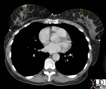
In this peri-menopausal woman the layer that houses the mammary apparatus has been almost entirely replaced by fat giving us an opportunity to image the surrounding capsule. It is best seen surrounding the mammary apparatus of the right breast. Can you see this structure? In the image below the outlines of the capsules have been interpolated. Courtesy Ashley Davidoff MD 43646
|
| Capsule |
|
In this CTscan the capsule of the mammary apparatus (middle layer of the hypodermis) has been drawn in. You had already reviewed this CT scan when we were discussing the case of mammary duct ectasia in the section on the ducts of the breast which is noted on the right. Courtesy Ashley Davidoff MD 43646b01
|
Applied Anatomy
Malignant disease that originates in the lobules and ductal system is considered “in situ” as long as the basement membrane does not get breached. The transgression of the basement membrane is an important microscopic and biological sign that the lesion has evolved into an aggressive entity and requires a plan for aggressive treatment. If not diagnosed at this point, a malignant lesion will progressively advance through the relatively weak caspular barriers of the lobe, mammary apparatus, and finally through the more resilient barrier of the skin, or posteriorly through the pectoralis fascia into the muscle. At this stage the cancer is classified as a stage III cancer. Stage I disease is the earliest stage with no spread of the disease, while stage IV is the most advanced disease, with widespread involvement of other organs beyond the breast.
|
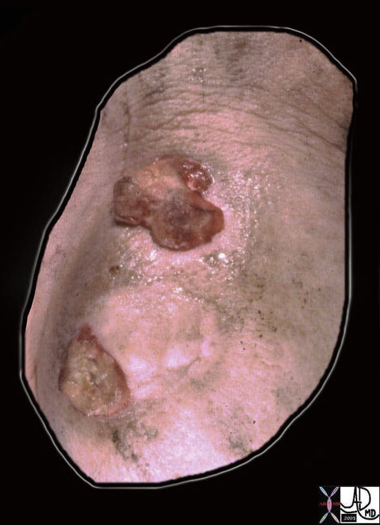
Tumor Breaching the Skin |
|
This gross pathology specimen is of a resected breast tumor as viewed from the skin surface. Two button size malignant lesions have breached the skin and advanced to the surface. Stage III is present. Courtesy Frank Reale MD 13526b04
|
| Tumor Breaching the Skin |
|
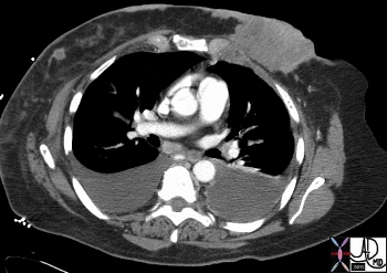
This CTscan through the chest shows a large left sided carcinoma that has breached the skin, and has spread along the skin toward the right breast, where a second nodule is noted. The primary lesion has also breached the pectoralis fascia and extended into the intercostal muscles. Bilateral pleural effusions with focal thickening of the left medial pleura suggest that there is systemic spread of the disease and stage IV breast carcinoma is most likely. Courtesy Ashley Davidoff MD. 43619
|
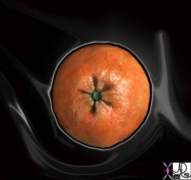
Peau d’orange (peel of an orange in French) is a clinical sign in which the skin of the breast is likened to the dimpled skin of an orange. In this entity the lymphatics are obstructed and the skin becomes edematous. As a result there is a dimpling of the skin.
| “Peau d’Orange” |
|
The peel of an orange or tangerine has natural dimpling with pits that are less than a millimeter in size. When breast carcinoma obstructs the lymphatics the edema that results causes a dimpling of the skin, much like the normal appearance of the skin of an orange. This sign has thus been called “peau d’orange” – the French for “skin of orange”. Courtesy Ashley Davidoff MD. 13575b02
|
Support Ligaments
Cooper’s ligaments run between lobes and lobules and span the breast from the corium of the skin to the fascia of the muscle. Cooper’s ligaments are composed of dense fibrous tissue.
They keep the breasts in their characteristic shape and position. In the elderly or in pregnancy these ligaments become loose, stretched, or may even fracture. As a result the breasts sag and they do not return to their former tension.
| Cooper’s Ligaments |
|
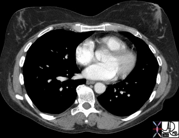
In this 58year old female there are fine strands of connective tissue connecting the mammary apparatus to the skin anteriorly. These are called Cooper’s ligaments. The more carefully you look the more you will see them. Their posterior attachments to the chest wall are not visible on this image. Courtesy Ashley Davidoff MD 43647
|
| Cooper’s Ligaments |
|
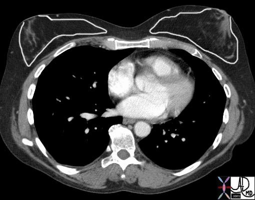
In this CT scan I have outlined the ligaments of Cooper in orange. Courtesy Ashley Davidoff MD 43647b03
|
| Cooper’s Ligaments |
|
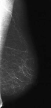
On mammography Cooper’s ligaments usually present as overlapping, criss-crossing, fine linear structures as seen in the overlay. Courtesy Priscilla Slanetz MD MPH 42873
|
Connective Tissues and Supporting Structures
Cooper’s ligaments run between lobes and lobules and span the breast from the corium of the skin to the fascia of the muscle. Cooper’s ligaments are composed of dense fibrous tissue.
They keep the breasts in their characteristic shape and position. In the elderly or in pregnancy these ligaments become loose, stretched, or may even fracture. As a result the breasts sag and they do not return to their former tension.
| Cooper’s Ligaments |
|
In this 58year old female there are fine strands of connective tissue connecting the mammary apparatus to the skin anteriorly. These are called Cooper’s ligaments. The more carefully you look the more you will see them. Their posterior attachments to the chest wall are not visible on this image. Courtesy Ashley Davidoff MD 43647
|
| Cooper’s Ligaments |
|
In this CT scan I have outlined the ligaments of Cooper in orange. Courtesy Ashley Davidoff MD 43647b03
|
| Cooper’s Ligaments |
|
On mammography Cooper’s ligaments usually present as overlapping, criss-crossing, fine linear structures as seen in the overlay. Courtesy Priscilla Slanetz MD MPH 42873
|
|