
Cancer
Common Breast Diseases
Malignant breast disease
Most diseases both benign and malignant arise from this highly active terminal ductal lobular unit (TDLU) in which there is constant change of structure. (Stirling)
The carcinomas are the most significant lesions of the breast. There are two major types of breast carcinoma that arise from the lobule namely ductal carcinoma and lobular carcinoma. They both arise from the TDLU.
Carcinoma of the breast at this level is divided into the non invasive or “in situ” carcinomas on the one hand and the invasive carcinomas on the other. The “in situ” carcinomas are restricted to the lobule and do not show invasion of the basement membrane while in the invasive group, there is obvious transgression of the basement membrane.
The “in situ” group has variable behavioral patterns and outcomes. These lesions may remain dormant for a lifetime, or may even involute. They do however place the woman at increased risk of developing cancer and should therefore be treated with heightened awareness.
The two types of “in situ” carcinomas are called ductal carcinoma in situ (DCIS), and lobular carcinoma in situ. (LCIS) and they are very different. DCIS is common and occurs in 70-95% of women at autopsy and is commonly associated with microcalcifications on mammography. It arises from ductal epithelium. LCIS is less common, clinically and radiologically more elusive, occurs in 1-5% of biopsies, and is rarely associated with microcalcifications on mammography. Wellings Once LCIS is identified the risk of developing cancer if untreated is about 30% within 20 years of diagnosis. It is not considered a precancerous disease, but a marker of increased risk.
The invasive carcinomas of the breast are correspondingly called invasive ductal carcinoma and invasive lobular carcinoma and are defined by the transgression of the basement membrane by malignant cells.
The most common site of involvement is the upper outer quadrant since as we have learned it is the location of “extra” tissue called the axillary tail of Spence. The presence of a larger amount of glandular tissue raises the relative incidence of disease in this quadrant. About 45% of carcinomas arise from this upper outer quadrant.
Invasive ductal carcinoma is the most common type of carcinoma and it has a tendency to incite a fibrous response which has structural consequences. Firstly fibrous tissue is very dense so that this type of tumor has a relatively dense appearance on mammography. The scirrhous and desmoplastic nature of fibrosis also causes a tugging effect on other structures such as the Cooper’s ligament, the skin or the nipple so that retraction of these structures can occur. Peau d’orange is the dimpling of the skin in carcinoma that is reminiscent of the dimpling seen in the skin of an orange, and it is caused by the effect of the carcinoma on the ligaments of Cooper. New onset of nipple retraction is also an important warning sign of cancer.
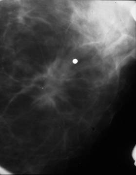
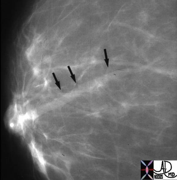
This mammogram shows a spiculated mass in the center of the image just rightward of the marker, and was shown to be an invasive ductal carcinoma. Courtesy Priscilla Slanetz MD MPH 42932 TheCommonVein.net
Fibrocystic disease
Fibrocystic changes in the breast are the most common entity of the breast and accounts for the common lumpy bumpy feel of normal breast tissue. It is a benign entity that arises from the TDLU. The pathology is caused by proliferation of the connective tissue and or epithelial tissue, resulting in cystic change and or fibrous changes. It has a variety of clinical, imaging and pathological variations including a single dominant cyst, diffuse irregularity caused by fibrocystic change and diffuse irregularity with a dominant lesion (U Virginia) The entity is subject to morphologic changes with hormonal changes of the menstrual cycle, and it may be associated with pain. The lesion is most common in the upper outer quadrant where the lobules are more voluminous in the tail of Spence.
Fibroadenoma
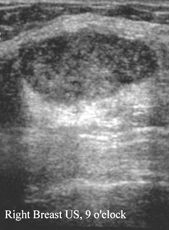
Fibroadenoma is a common benign disorder of younger adult women that arises from the intralobar connective tissue elements and from epithelial elements. Unlike the cyst above the fibroadenoma presents on ultrasound as a well circumscribed solid lesion.
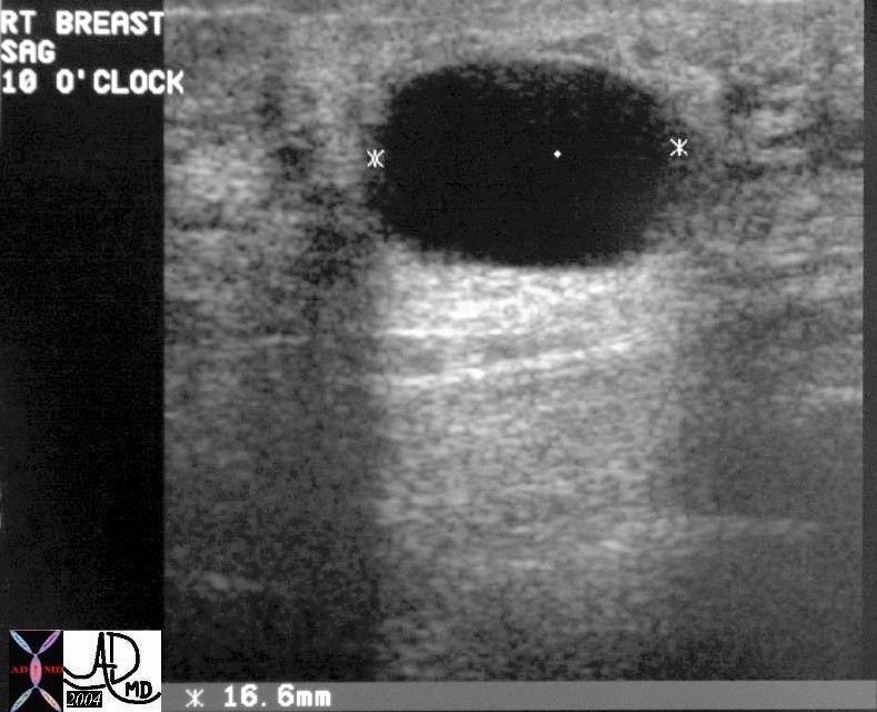
This ultrasound of the left breast of a young adult female reveals a well circumscribed mass that proved to be a fibroadenoma. Courtesy Priscilla Slanetz MD MPH TheCommonVein.net 42714
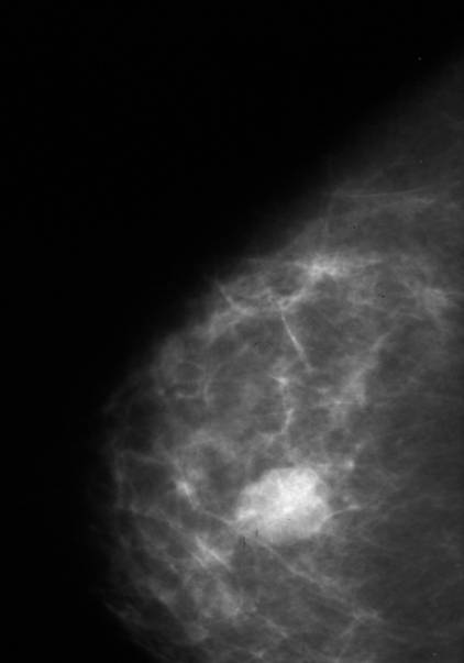
This mammogram of the left breast of a young adult female reveals a well circumscribed mass that proved to be a fibroadenoma. Courtesy Priscilla Slanetz MD MPH TheCommonVein.net 42913
Diseases of the ducts
Disease of the larger ducts is relatively uncommon. Duct ectasia is a dilatation of the major ducts just behind the nipple that occurs in women nearing menopause and is really a normal finding. Accumulation of secretions may cause blockage of the duct which can then become symptomatic.
This 46year old female demonstrates prominent tubular structures near the nipple of the left breast. This image was previously used to demonstrate the ducts. A diagnosis of duct ectasia is most likely. Courtesy Ashley Davidoff MD TheCommonVein.net 43646
This mammogram of the right breast shows a dilated duct coursing toward the nipple. This is the mammographic finding of duct ectasia. Courtesy Priscilla Slanetz MD MPH TheCommonVein.net 13563
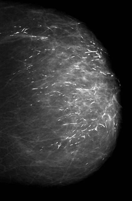
The mammogram of the right breast shows benign tubular calcifications all oriented in the same way toward the nipple. The calcifications are characteristic of benign secretory calcifications within the ducts. Courtesy Priscilla Slanetz MD MPH TheCommonVein.net 42885
Nipple discharge
Nipple discharge is the third most common breast symptom after mass, and pain of the breast. Discharge is a normal event in women who are in the reproductive age. 50-60% of women can express some secretion on self examination. It is uncommon that a discharge is associated with breast cancer. Causes of nipple discharge include persistent secretion after pregnancy, benign hormonal affects, and mammary duct ectasia. Phenothiazines may also cause a nipple discharge. Benign features include bilaterality, and the involvement of multiple orifices. Bloody discharge that is spontaneous, from a single duct should raise concern for malignancy, but even so, only 10% of these patients have an underlying malignancy. Intraductal papilloma is the most common cause.
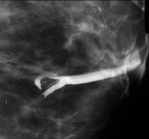
This is a case of a 48 year old female with bloody nipple discharge. Ductogram reveals a segmental lactiferous duct with filling defect consistent with a diagnosis of intraductal papilloma. Courtesy Priscilla Slanetz MD. MPH TheCommonVein.net 43030
This is the correlative ultrasound of the above case. Note the dilated duct within which resides the echogenic nodule. Courtesy Priscilla Slanetz MD. MPH TheCommonVein.net 43031
Pagets’s Disease of the Nipple
Paget’s disease of the nipple is a rare disease of the nipple characterized by eczematous change of the nipple and almost always associated with an underlying breast carcinoma, specifically with high grade DCIS. The areola may also show eczematous change. It occurs in about 12% of patients with breast carcinoma. The findings in the nipple may be the only indication of disease, and so a new scaliness of the nipple warrants serious attention of clinician, mammography technologist, and radiologist. Only about 50% of patients with Paget’s have a radiographic abnormality on mammography.
Infection
keywords breast skin prosthesis fx air dx abscess CTscan C- Ashley Davidoff TheCommonVein.net
22983