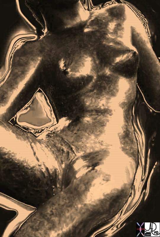
The Life Cycle of the Breast
Copyright 2010
About the Exhibit
The Life Cycle of the Breast is a tribute to the inner beauty of the breast from its cellular makeup, to the special tissues that contribute to its structure and function, and particulalrly the mechanism allowing the source of nutrition for the infant child.
The exhibit was created in part, from a larger educational exhibit presented at the international meeting of radiologists in Chicago – Radiologic Society of North America and has been embellished to reflect the a cycle of the breast from the time of conception when the genetic material is handed over through to return . It is part of an educational program for physicians called The Common Vein and is being exhibited at the Newton Open Studios in May 2010.
Purpose of the Exhibit
The purpose of the exhibit is to promote understanding of the structure and function of the breast in health and disease and to express the inner beauty of the breast through artistic medium.
Introduction
The coordinated function of the cells and ductal systems eventually mature into a lactating organ. The primary function of the organ only exists for a very small portion of the life of child bearing women. For the rest of adult life, the beauty of the breast as a secondary sex organ fills the minds and imaginations of men and women alike with all sorts of fantasies.
The first part of this exhibit is dedicated to the beauty of the biology of the breast , and the second is dedicated to breast cancer.
About the Artist
Dr. Davidoff’s interests in biology, nature and creative arts converge in The Life Cycle of the Breast. Conventional medical images are augmented and artistically rendered to portray medical and philosophical concepts and to highlight the beauty of the human female breast. These works present the breast from the time of conception, through embryonic development, birth, growth, aging, disease and return, highlighting its special aesthetics during prime function, and then the story of cancer .
Biographical Statement
Ashley Davidoff is Clinical Professor of Radiology at the University of Massachusetts and a clinical radiologist at Caritas St Elizabeth’s Medical Center. He received his medical training at the University of Witwatersrand in Johannesburg, South Africa. After immigrating to North America he was credentialed in pediatric cardiology at The Hospital for Sick Children, University of Toronto. Subsequently he trained at two Harvard Medical School affiliates, specializing in pediatric cardiac pathology at Children’s Hospital in Boston, and then completing radiology residency and cardiovascular fellowship at the Brigham and Women’s Hospital. Dr. Davidoff plays an instrumental role in medical education, having won numerous teaching awards and directed several regional medical education courses. His award winning modules in medical education have been exhibited at national radiology meetings.
Dr. Davidoff combines his professional medical interests with artistic endeavors, focusing on form and function in nature. His artwork portrays and incorporates structure of the body and structure in nature in a variety of media. His photography and digital art renderings are currently on exhibit at the Museum of Science, and his sculptural pieces produced from natural elements are displayed in the annual Newton Open Studios exhibit, in his award winning garden. He is happily married to Naomi Fisher and they have four wonderful children, Talya, Arielle, Aaron, and Daniella.
Address: 3 Lake Avenue, Newton Center 02459
Email: ad@thecommonvein.com
Web Site: thecommonvein.com
|
Conception The Sperm, The Egg, and the Spark of Life |
|
The meeting of a single sperm with a single egg creates and instills a spark that fires the soul. An inexplicable inspiration gives birth to a new and unique life. The background greens and blues represent the raw materials of heaven and earth. Davidoff art copyright 2008 13275a12.800
|
Development
While in the uterus, the primary glandular breast tissue develops from primitive tissues. All the building blocks that are requiresd for the mature breast are present in utero. They consist of the skin, glandular tissue, connective tissue and adipose tissue, as well as the blood vessels, nerves, and ducts. In fact, once the building blocks are formed, they become subject to the effects of the mother’s hormones that are circulating in her blood through the placenta.
Development Raw Materials Multiplication Differentiation connection to the Mother
|
| 13275b01i02e02 fetus raw material amniotic cavity Cycles of differentiation Davidoff art copyright 2009 |
|
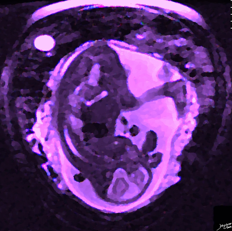
The Female Fetus in a World of Her Own |
|
When the building blocks of the breasts are developed they are subject to the effects of the maternal hormones. The image was created from an MRI, and the image rendered to create an artistic effect and reflect the female status 46701b01.4k.8s MRI T2 weighted Davidoff MD Davidoff Art Copyright 2009 all rights reserved |
A Short Essay
The times and cycles of my life – A story of womanly issues
A Short Essay in Poetic Form copyright 2010
Background
The following short story written in poetic form, refelects a personification of the breast tissue in the embryo. The building blocks of the breast develop from primitive embryonic tissue called mesoderm (mesenchyme) and ectoderm and once formed are subject to the effects of the mother’s circulating hormones. The breast tissue of the developing fetus is thus stimulated during pregnancy and in the early days after birth, while the maternal hormones are still present . The newborn may in fact have some prominent breast tissue and also produce some secretions during this time. As the maternal hormones in the newborn metabolise in the post partum period and are not replenished, the breast tissue regresses and secretions cease.
The Story
I was born of the blanket of the flesh that covered her chest. Although I knew my mother I had no clue who my father was.
I was barely three months old. A lot of things were happening around me. Cells and tissues were scurrying around in a hub of construction. Time seemed to be moving fast and a lot of refinement of structure was evolving on the already well established structural foundation. After a brief consideration of taking a breather I suddenly got a new energy and a yearning to explore … there seemed to be something missing from my life – I needed fulfillment – I needed to become whole.
Aside from the sounds of ebb and flow and a beating heartbeat from the core, there were no other sounds. Nobody said a word. It was if all they all knew exactly what to do and were driven by a force greater than themselves. This was quite frightening but the organization seemed so magnificent and the vision of this integrated body seemed so noble that I followed my instincts in similar silence. When there was some respite of activity, silent messages were transmitted through the cell vine. The vision was to build a human force that not only had body but also had mind and soul. Well those too sounded noble but I had no clue what they might mean. I nodded in affirmation since this seemed to be the gesture of all and I hoped that someday I would understand – blind faith I suppose.
Well back to this incomplete feeling and my yearning for fulfillment. It seemed that I did not have what it would take to play any part in the grand person we were constructing. I felt almost as if part of my body was missing.
Without conscious decision I suddenly found myself hurtling through the dermis into the hypodermis with a destiny that seemed like an inextricable link with the Miss Mesenchyme who came from matriarch Leah, the mother of vessels, muscle, and connectivity of all tissues. Leah’s functtion in life was bringing us all together. During my unintentional journey toward Miss Mesenchyme the union to my surprise seemed like a pleasurable destiny that was an absolute for my completion as a biological entity. It was not a matter of a love affair or anything like that. It seemed that for my wholeness, I absolutely needed to combine forces with Miss Mesenchyme who was heading toward me. The forces of our union were beyond our control … and now we were one and our destiny we were told was as important as any. We would be the fuel of future generations. Babies would lust after us and were going to drink the fruit of our factories with gusto. Men would bow down to us. Industry would revel in our form and build billions of brassieres to support us – we had no idea what this was all about. Anyway together we felt important and good about ourselves.
What did we look like at this stage? In a word we were scrawny. We certainly did not look like the heroines we were promised to be. We could be described as long and lanky tubes– tree like in our branching pattern – winter trees that is.. We were dormant without leaves and without function. We felt naked without leaves and without the promised cushions of fat that were to come later in life. We consisted at this stage of 18 tubes radially distributed around the nipple. In the far distance I could see the ocean or so it seemed like an ocean. I peaked out and met Ms Areola and Ms Nipple as well – all really part of me.
And then suddenly there was the “jolt” almost like a “bolt” of lightning. Our rapidly evolving mistress was growing in leaps and bounds but she was far from the goddess of body mind and soul. And then I felt a rumbling as my engines started to fire up. I knew we were getting close to delivery but I was only going to be a baby – nowhere nearly able to nurse . So why were my engines starting I said to myself?
The bloody ol Estra Jen – some called her Esther’s gin – because she was a slave driver and could drive us to drink When she is around it is work work work every minute and hour Where did this Estra Jen come from? – My ovaries were certainly not working yet. I looked down – and yes certainly – quiet as a dormouse – our family jewels – resting. I did not understand the forces around and beyond me, but I had faith that I was in for an awesome ride.
Principles of the Body – Principles of Society
There are basic principles of structure and function, that apply to all the organs in the body.
The body is made up of biologic units – or building blocks, that work in concert for the benefit of the whole. Each unit, be it a cell or an organ in the body is valuable because it is able to receive a raw product which with its functionality is able to process and create a new valuable product, which it exports to the community at large to help the body survive and thrive. The body like our society is made of institutions or factories or cells of functionality and productivity, that are valuable to the society and they need transport systems to move things and they need a controlling system to make sure that supply meets demand.
There is a remarkable consistency of the structure of the atom and the solar system with a central nucleus and revolving smaller body bound to the nucleus by physical forces. Biology with all its mysteries is made up of those atoms, that join together to form more complex structures with macromolecules forming into organelles finally being “provided” with a life force in the cell which is the most basic biological unit. The cell has structure and function at a simple level but as it joins with other cells of a similar ilk they form a new structure more powerful than the individual parts – the organ. This pattern of units to unity where 1+1= ONE where one is a new unit bigger and more powerful than the individual parts is a recurring pattern extending from the organ to body systems – to the person, the couple, the family, and to the community at large.
From the Atom to the Solar Sytem with biology in Between |
| 13440c11.8 units to unity atom elements molecules chromosomes cells tissues organs systems body part abdomen chest person couple family home community village city state country worls solar system Davidoff art copyright 2008 |
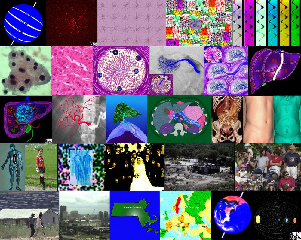
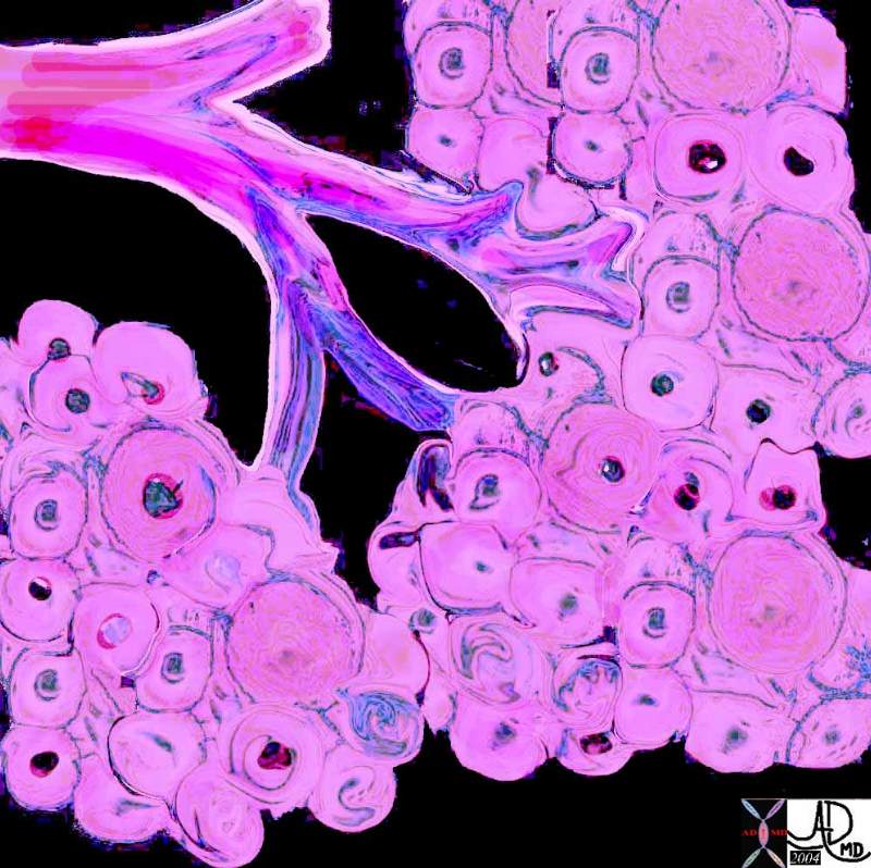
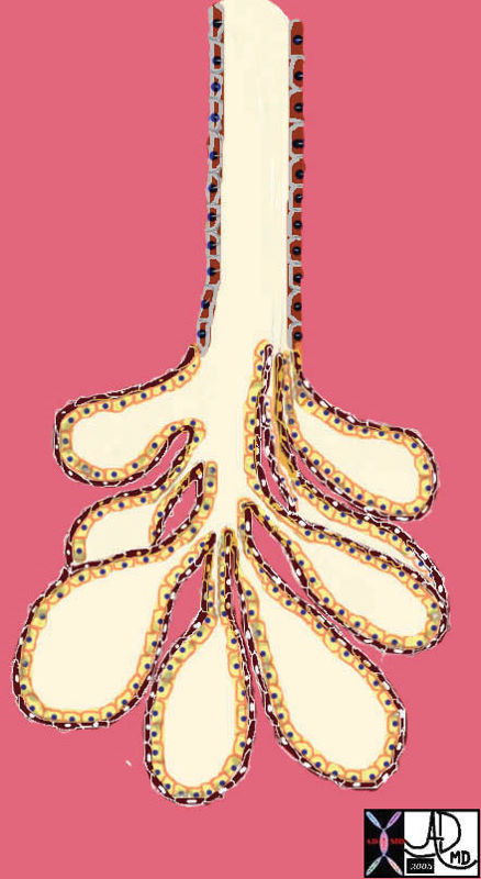
The Glandular Tissue
The glandular tissue becomes the important milk producing tissue and consists of cells organized in a rosette around a central duct. The cells will eventually produce milk and secrete the milk into a small duct. There are thousands of these systems in each of the breasts and all the small ducts join – like branches of a tree into bigger and bigger ducts culminating in 15-20 large lactiferous ducts that empty on to the nipple. These building blocks are present in the fetus. They can be considered small factories of production, and the ducts are the transport systems that take the milk to its final destination.
Cells and a Ductule of the Breast Factories of Production
|
|
The pink cells have a central blue nucleus in which the genetic material resides. Thousands of years of genetic history is present consisting of the genetic line as well as the biological history of the individual. The genetic material contains the recipes of how to make milk that will enable the baby to survive. The genes also contain both healthy news and potentially unhealthy news with some information that predicts events that will unfold for the individual. Current science is trying to read that history in order to predict the health and disease of the individual. 39939b03.800 exocrine gland epithelium ductule histology cytoplasmic granules nucleus Davidoff art |
This system, like all the systems in the body, is a micrososm of our society. In the same way that our society functions in the production of material with supply and demand forces, so the cells have to function in the same way. They eventually have to produce the milk in sufficient quantities for the need of the baby. Once produced, the milk needs to be transported to the customer who demands delivery in a timely fashion. The breast has to learn and know how to make the milk, understand the laws of economics of supply and demand and then know how to transport it so that it is farm fresh on delivery . In summary – the cells represent the farms and the factories of our cities and towns. The ducts represent the transport systems – highway byways trucks trains ships and planes that bring the product to the customer. Control mechanisms that coordinate the production and delivery, are the nerves, and hormones. All systems work the same way in the body and in society. They receive a raw product, with their abilities they process the product and they export the product. Their function needs to be controlled by a governing body who has a sense of supply, demand, and need.
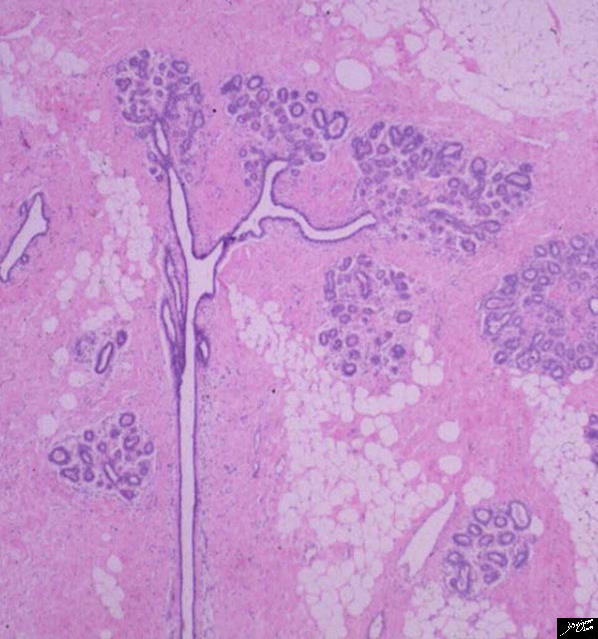
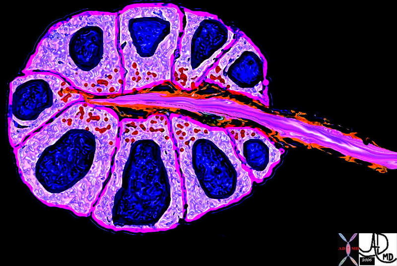
This image is an actual histological section of a normal adult breast as een under the microscope under low power. The tubular structures (white) subtending the small rosettes of glandular structures (purple) are central . The pink background is the actual color of the supporting connective tissue using hemotoxylin and eosin stain. The white “bubbles” represent fatty tissue. Note the tree like structure of the ducts and glands.
breast lobe lobule terminal duct lobular unit TDLU adipose tissue stroma connective tissue gland glandular tissue normal anatomy histology Courtesy Frank Reale MD TheCommonVein.net 13511d03s
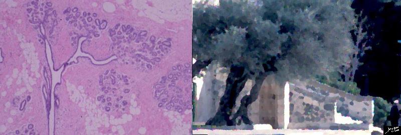
The tree like structure is universal in the body and universal in nature. The tree like glandular structure that is thousands of years old is put side by side with a rendered photograph of an olive tree in the gardens of Gethsemane in Israel where some of the olive trees are thousands of years old as well, but of course not as old nor as wise as the glandular tissue of the breast!
Courtesy Ashley Davidoff Courtesy Frank Reale MD TheCommonVein.net 13511d02.8s
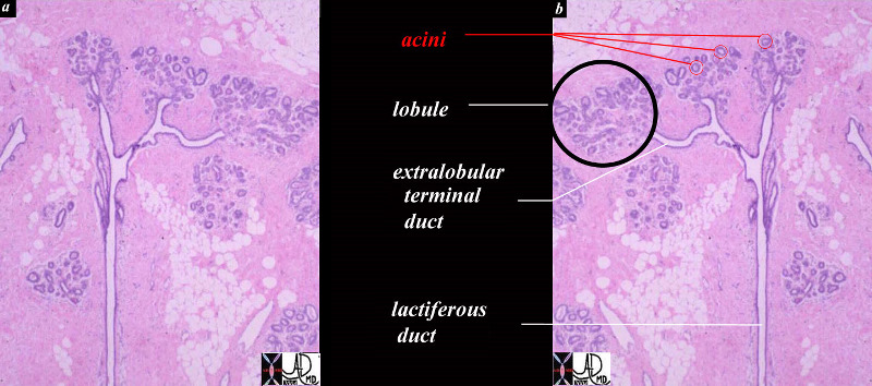
This diagram shows a terminal duct lobule unit. (TDLU) A lobule is ringed in black and the extralobular terminal duct is noted and labelled. The acini are shown ringed in red but the draining intralobular ducts are not seen The lobules and extralobular terminal ducts join to form a single lactiferous duct which exits at the nipple
Courtesy Frank Reale MD TheCommonVein.net 13511b01c05.8s
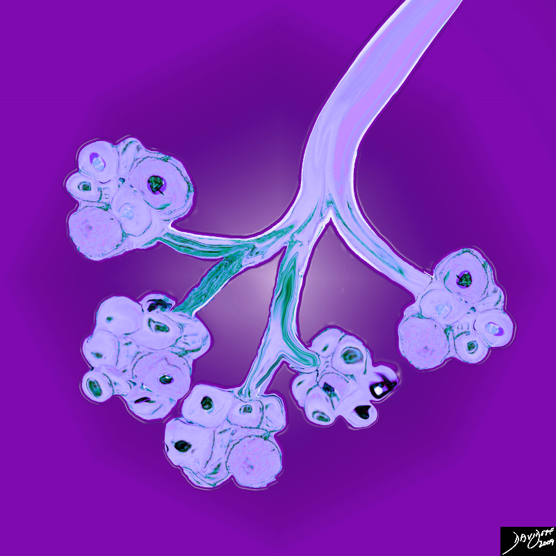
The Lobule Glands Acini and Ducts of the Breast Artistic View
|
| 32645a05b05b.8s breast acini terminal ductule intralobular terminal duct histology Davidoff art |
The Cells that Make up the Breast
|
42707b03b45b04 |
| 42707b03b45b04 breast mammary gland terminal duct lobular unit epithelium acinus acini myoepithelial cell duct ductule Courtesy Ashley Davidoff MD |
|
85198f00b.08ks |
| 85198f00b.08ks cells epithelium columnar epithelium nucleus cytoplasm cell basement membrane mucus submucosa muscularis serosa normal histology Davidoff art Courtesy Ashley Davidoff MD copyright 2009 |
|
85198k03.8s |
| 85198k03.8s cell normal columnar cell histolgy cytology Davidoff art copyright 2009 All rights reserved |
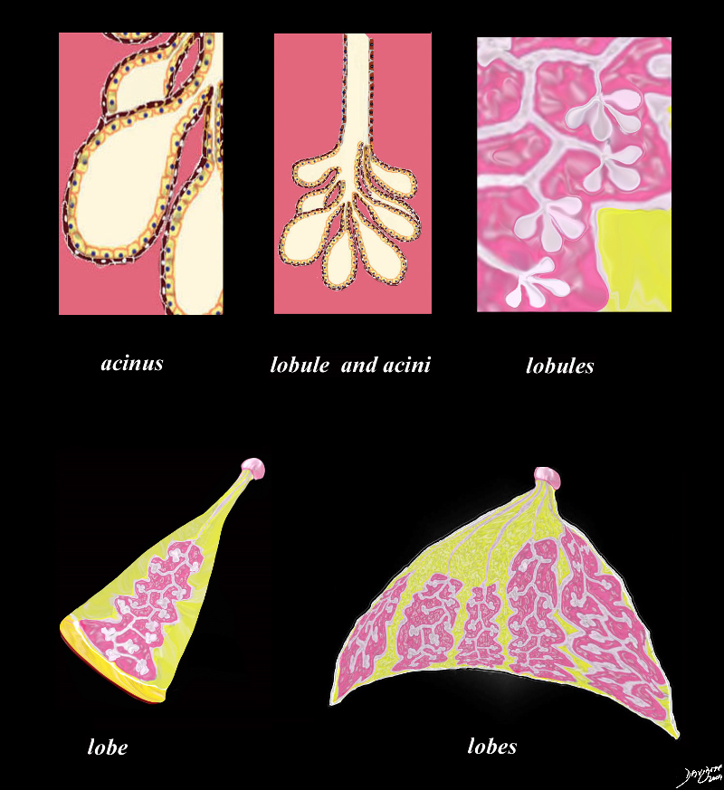
The Units that Make up the breast
Units to unity applies to all structures of the body The following is a description of the makeup of the breast.
Parts of the Breast Units to Unity |
| 42707c30.8s This drawing shows the part that make up the whole. The acinus is the single basic glandular unit and consists of an inner lining of cuboidal cells that rests on a thin muscle layer. The cells of the acinus secrete milk into the lumen of the gland which is transported into the tubular ductule. The lobule is made of a group of acini. Multiple lobules join together and form a lobe which is subtended by a duct which has an independent exit on the nipple. Finally 14-20 lobules combine to form the glandular tissue of the breast that is supported and padded by adipose tissue (fat) that is shown in yellow color. Each of the lobe is subtended by a single duct which empties onto the nipple. code breast normal anatomy histology lobe lobules acini alveoli acinus lactiferous duct extralobular ductule intralobular ductule Davidoff art Courtesy Ashley Davidoff MD copyright 2009 all rights reserved TheCommonVein.net |
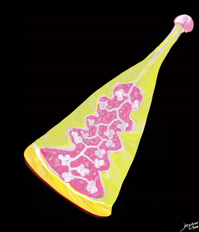
The Lobe of the Breast |
| 42707b03b38b03.8s breast mammary gland lobe lobule adipose tissue layers fat nipple interlobular fat adipose tissue stroma connective tissue parenchyma glandular apparatus mammary apparatus lactiferous ducts ductules acinus acini normal anatomy drawing Courtesy Ashley Davidoff MD TheCommonVein.net |
18-20 Lobules Form the Glandular Tissue of Each Breast |
| 42707b03b29b03 CC view breast mammary gland nipple adipose tissue layers fat superficial adipose tissue deep retromammary interlobular fat adipose tissue stroma connective tissue parenchyma glandular apparatus mammary apparatus lactiferous ducts ductules acinus acini normal anatomy drawing Courtesy Ashley Davidoff MD TheCommonVein.net |
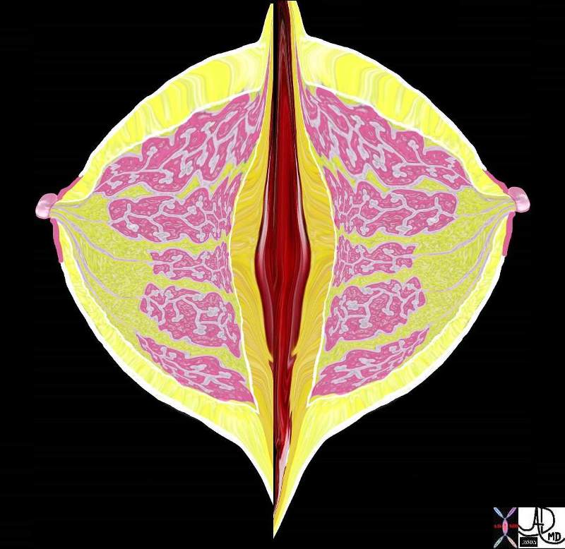
Tree Like Form of the Glandular part of the Breast
The breast
Artistic Rendition of the CTscan of a Breast |
| 46792b09.800 breast glandular tissue applied biology applied anatomy Davidoff art Davidoff trees TheCommonVein.net |
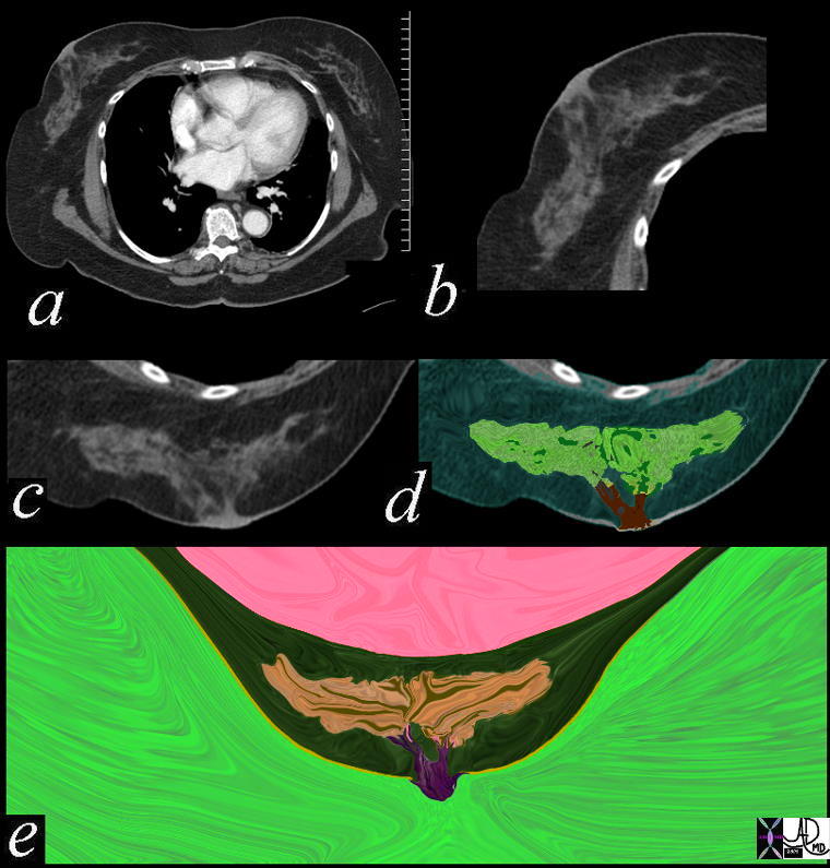
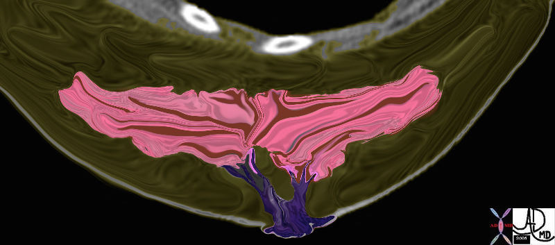
Tree Like Structure in the Breast CT Scan |
| 46791c02b01.800 breast glandular tissue applied biology appliued anatomy Davidoff art Davidoff trees TheCommonVein.net |
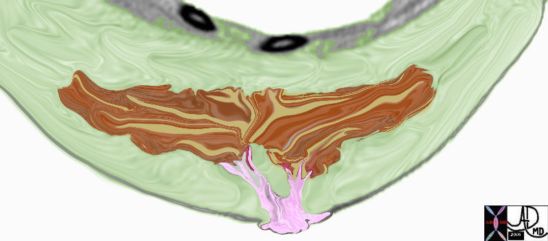
46792b12.800 |
| 46792b12.800 breast glandular tissue applied biology appliued anatomy Davidoff art Davidoff trees TheCommonVein.net |
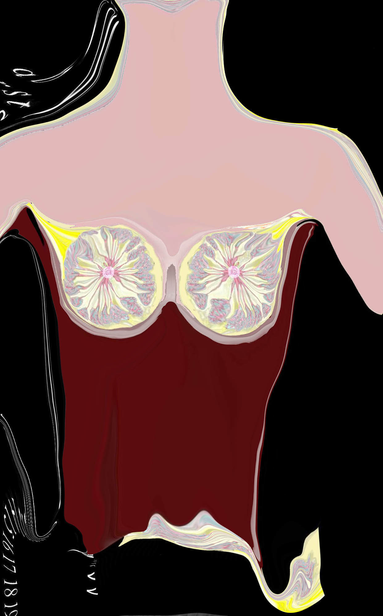
Skin Deep Artistic Rendition of the Histology of the Breast |
| 42657b15 Courtesy Ashley Davidoff MD breast lobule duct nipple fat glands glandular tissue normal anatomy drawing art Davidoff art |
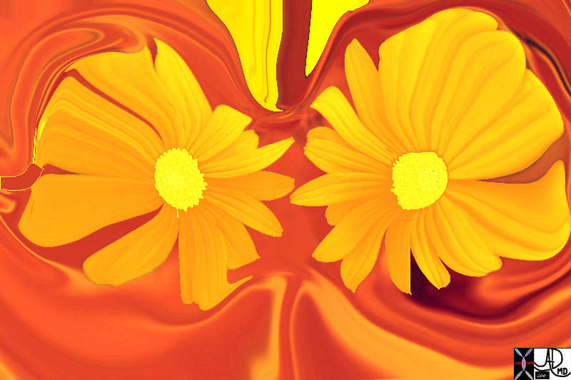
Artistic Rendition |
| 79560p.800 flowers breasts structure anatomy Davidoff art Davidoff photography TCV applied biology The Common |
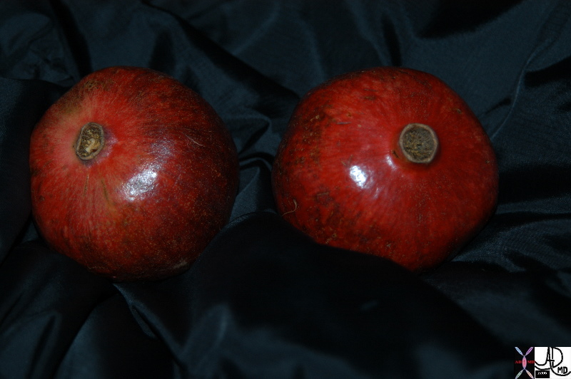
Artistic Rendition |
| 86226.8 pomegranates breast red nipple food in the body food biology medicine and body fruit Davidoff art Davidoff photography |
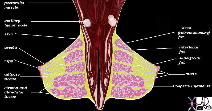
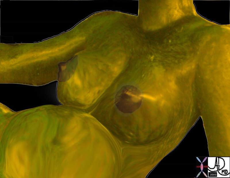
Anatomy
|
42707b03b25bL |
| 42707b03b25bL breast mammary gland nipple areola ducts Coopers ligaments Cooper’s ligaments pectoralis muscle chest wall adipose tissue layers fat superficial adipose tissue deep retromammary interlobular fat adipose tissue stroma connective tissue parenchyma glandular apparatus mammary apparatus axilla axillary lymph node normal anatomy drawing Courtesy Ashley Davidoff MD |
|
78378pb06 |
| 78378pb06 breast young normal anatomy sculpture DB |
78378pb06l03.8s |
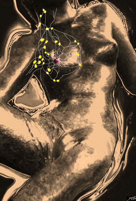
| 78378pb06l03.8s breast mammary gland lymphatic drainage lymph nodes axillary lymph nodes infraclavicular lymph nodes supralavicular lymph nodes internal mammary lymph nodes parasternal lymph nodes anatomy normal Courtesy Ashley Davidoff MD |
78378pb09a02 |
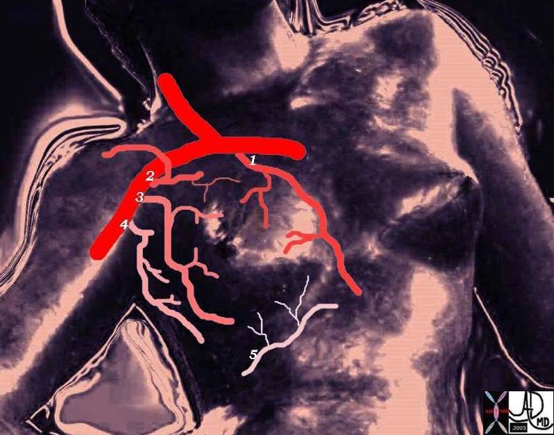
| 78378pb09a02 breast blood supply artery arteries subclavian internal mammary artery IMA perforators thoracoacromial artery thoracodorsal artery axillary artery lateral thoracic artery intercostal artery art normal anatomy Courtesy Ashley Davidoff MD |
78378pb09a03b |
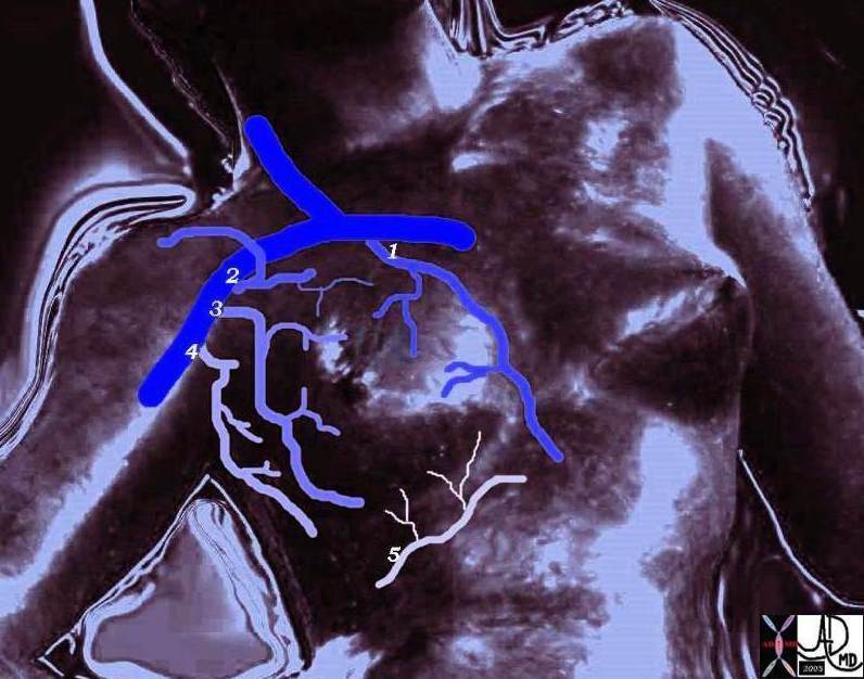
| 78378pb09a03b breast venous drainage vein subclavian internal mammary vein internal thoracic vein perforators thoracoacromial vein thoracodorsal vein axillary vein lateral thoracic vein intercostal vein art normal anatomy Courtesy Ashley Davidoff MD |
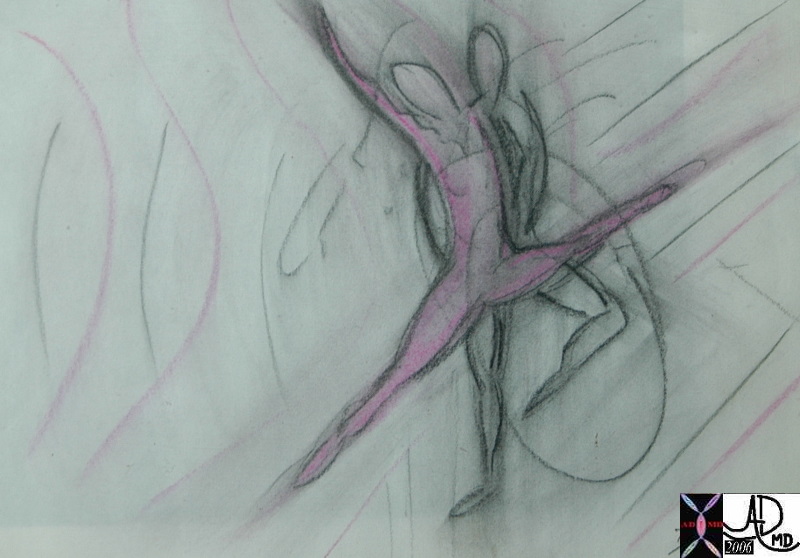
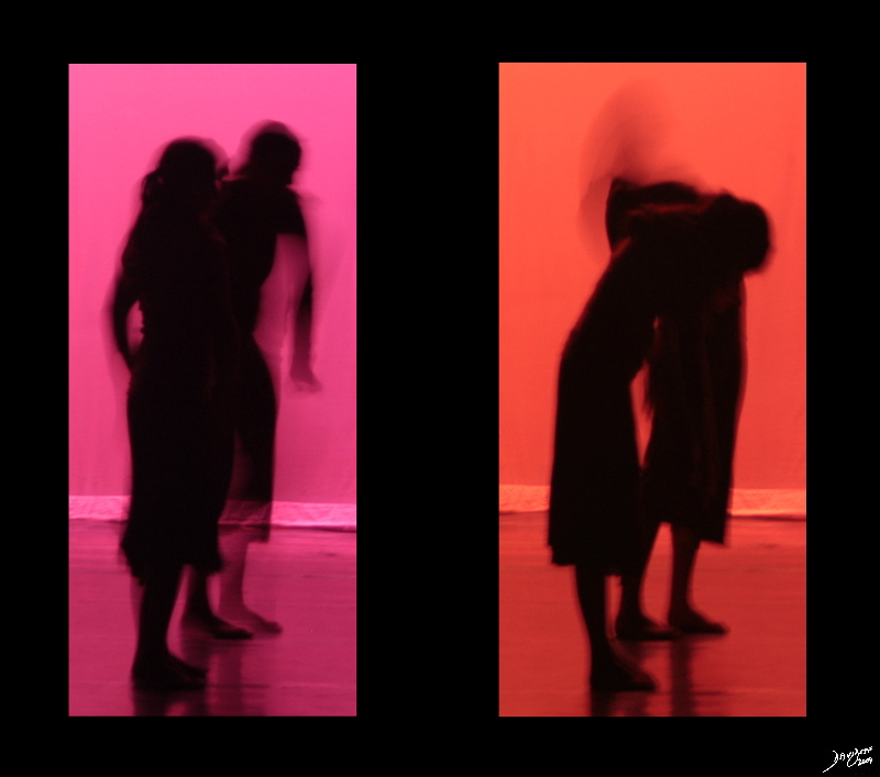
Feminity and Society
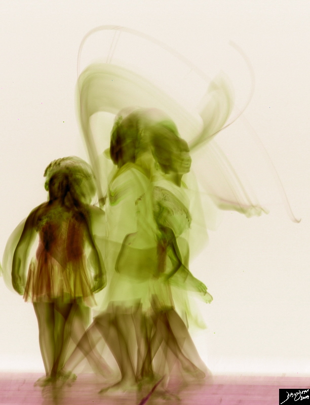
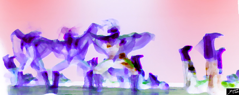
Growing Up – Changing Physiology |
| 86937pb01.8s dance movement Davidoff art Davidoff photography copyright 2009 all rights reserved TheCommonVein.net |
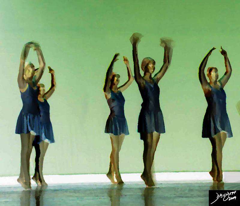
Not Quite There – Adolescence |
| 87103pb03.8k.8s dance movement units to unity individual uniqueness group Davidoff art Davidoff photography copyright 2009 all rights reserved TheCommonVein.net |
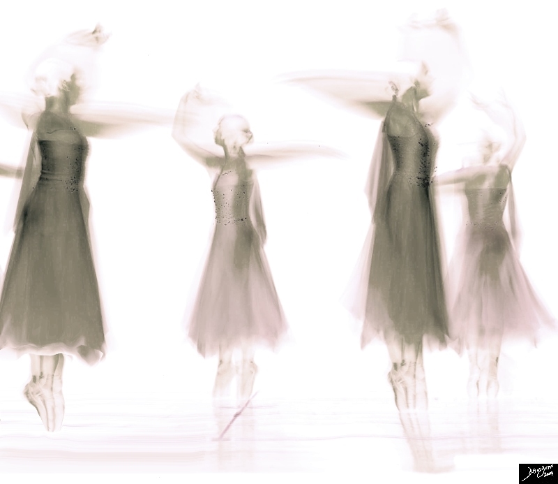
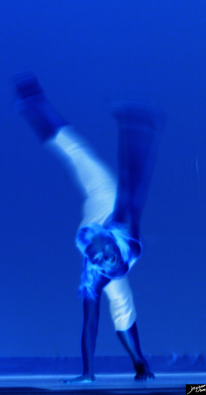
Soft Strong |
| 71919pb01.8s dance excercise movement spine lumbar spine thoracic spine balance muscles expression creativity cerebellum equilibrium middle ear extension extend point pointe Davidoff art Davidoff photography copyright 2009 all rights reserved |
Female Profile |
| 86995pb02.65r dance ballet Rosita’s dance portfolio Davidoff art Davidoff photography copyright 2009 all rights reserved TheCommonVein.net |
82730p |
| 82730p Duet Davidoff art Davidoff painting Davidoff drawing v |
Grace and Power in Blue Another Side Another Mood |
| 71900pb02.8s Davidoff art Davidoff photography copyright 2009 all rights reserved TheCommonVein.net |
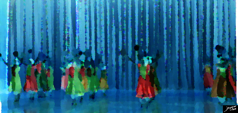
Another Culture |
| 94726pb.8s tambourine dance in blue dance Davidoff art Davidoff photography copyright 2010 TheCommonVein.net |
Physiology
Pregnancy
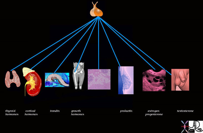
During pregnancy the breasts increase significantly in size, increasing by about 400grams per breast, under the influence of estrogen progesterone growth hormone insulin and cortisol. Prolactin also plays a role of breast enlargement during pregnancy resulting in an overall average increase in size of about 145mls. The higher levels of estrogen result in progressive proliferation, elongation, and dilatation of the ductal system while progesterone is mostly responsible for glandular development. Most of the fat of the breast is replaced in late pregnancy by enlarging glandular tissue necessary to produce milk. The nipple and areola become deeply pigmented and increase in size as well.
Following birth and delivery of the placenta there is a sudden drop of estrogen and progesterone levels. Prolactin plays an increasing role in breast function and morphology as milk production is initiated and continued throughout the nursing period.
|
Pregnancy |
|
Enlargement of the breasts and darkening of the nipples is the physiological response to pregnancy. The real physiological function of the breasts is limited to the short period in a woman’s life for breast feeding. 78380pb05b04 breast mammary gland pregnancy lactation anatomy normal art Courtesy Ashley Davidoff MD TheCommonVein.net |
|
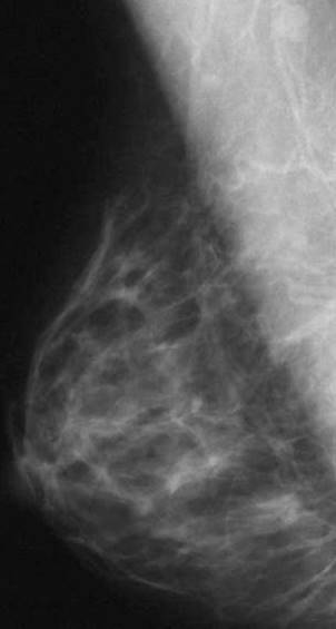
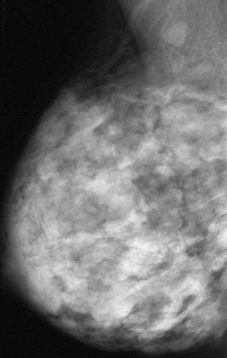
Non Lactating and Lactating Breast |
|
The first image is from a screening mammogram and the second image of the same patient was taken during lactation. Note the change in size and contours of the breast and how the predominant linear pattern of the non lactating breast becomes nodular and lobular with lactation. Courtesy Priscilla Slanetz MD MPH 43064b01 43065b01 TheCommonVein.net |
Non Lactating Glands and Lactating Glands |
| Courtesy Ashley Davidoff MD pancreas exocrine cell lobule acinus duct ductule normal histology parts drawing tube gland 32645a05b05b.8s 32645a06 TheCommonVein.net |
|
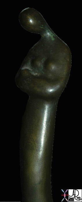
Infant and Mother |
|
The abstract sculpture shows the softness and tenderness of breastfeeding and motherhood highlighted by the postion of the mothers head and her arms. The sculptor is unknown and the sculpture is the property of the author 79392pb03 breast anatomy normal lactation sculpture Courtesy Ashley Davidoff MD TheCommonVein.net |
|
Lactating Breast |
|
The mammogram was taken during lactation and the technique requires compression of the breast. A small drop of milk ios seen art the lower edge of the nipple 02886b05 breast pregnancy lactation lactating female mammogram mammography Xerography Xerogram Courtesy Ashley Davidoff MD anatomy normal physiology TheCommonVein.net |
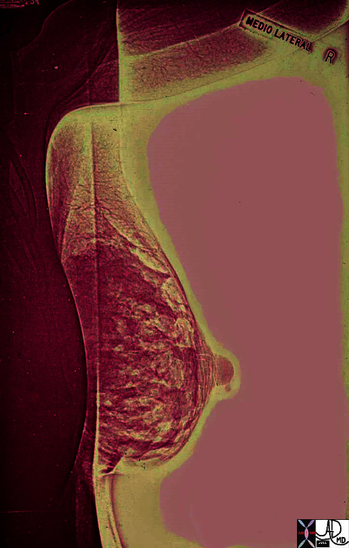
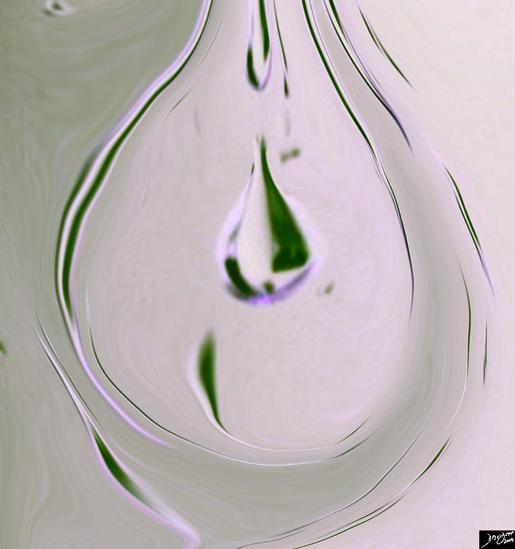
02504pb04d.8s drop milk lactation nursing breast Davidoff art TheCommonVein.net
As the infant starts to suck on the nipple a reflex production of prolactin from the anterior pituitary gland occurs. The prolactin in turn stimulates a cascade of hormonal events with a subsequent cascade of flowing milk. As stated the ducts of the apocrine glands act as storage sites for the milk produced by the acini. The breasts, are larger by about 10-20% as they become distended with milk (Dewey) In the first two days post partum there is only about 50ccs per day of milk produced, which increases dramatically to 500ccs per day by day 4. At 3 months post partum this usually plateaus at 850-1000ccs per day. If the mother feeds every 4 hours then at peak production prior to feeding there should be about 120ccs of milk in the breasts equating to about 50-60ccs per breast. (related links) The rate of milk production has been estimated at between 10-60ccs per hour (Daly) The basic size of the breast has no bearing on the volume of milk. At the peak of lactation the breasts become almost double their baseline size.
|
Control of Lactation by the Pituitary Gland |
| 72353 pituitary gland posterior pituitary gland breast lactation prolactin ovary estrogen FSH Davidoff art Davidoff drawing Davidoff MD TheCommonVein.net |
At a moment in time…
All is well and the community of cells within the breast are living happily and health
42707b03b45b04 |
| 42707b03b45b04 breast mammary gland terminal duct lobular unit epithelium acinus acini myoepithelial cell duct ductule Courtesy Ashley Davidoff MD TheCommonVein.net |
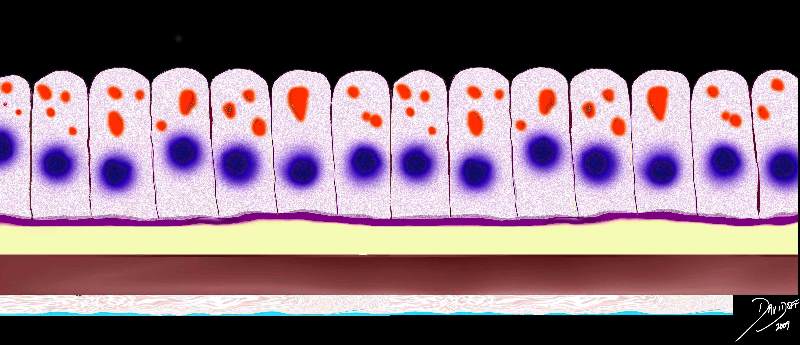
85198f00b.08ks |
| 85198f00b.08ks cells epithelium columnar epithelium nucleus cytoplasm cell basement membrane mucus submucosa muscularis serosa normal histology Davidoff art Courtesy Ashley Davidoff MD TheCommonVein.net |
85198k03.8s |
| 85198k03.8s cell normal columnar cell histolgy cytology Davidoff art copyright 2009 All rights reserved TheCommonVein.net |
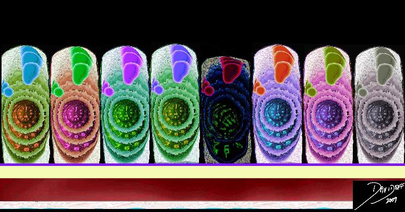
At another moment in time
during adulthood … for one of many potential reasons.. occasionally genetic, sometimes environment, and mostly unknown one of the cells lining the glands or ducts loses sense of time.. and as a result rebels against the community
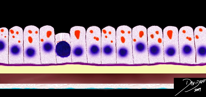
A Rabble Rouser rebel Arises in the Community of cells – Easy to Spot |
| 85198f01.08s cells epithelium columnar epithelium nucleus cytoplasm cell basement membrane mucus submucosa muscularis serosa histopathology Davidoff art Courtesy Ashley Davidoff MD TheCommonVein.net |
Sense of Time is Aberrant |
| 85198gc25.08 cells epithelium columnar epithelium nucleus cytoplasm cell time adenocarcinoma aberrant growth malignant cancer neoplasm death cycle normal life span age growth mucus dysplasia histopathology hyperchromatic nucleus increased nuclear to cytoplasmic ratio Davidoff art Courtesy Ashley Davidoff MD copyright TheCommonVein.net |
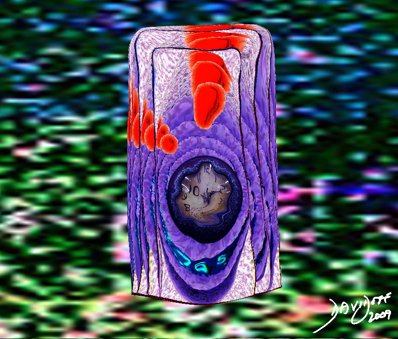
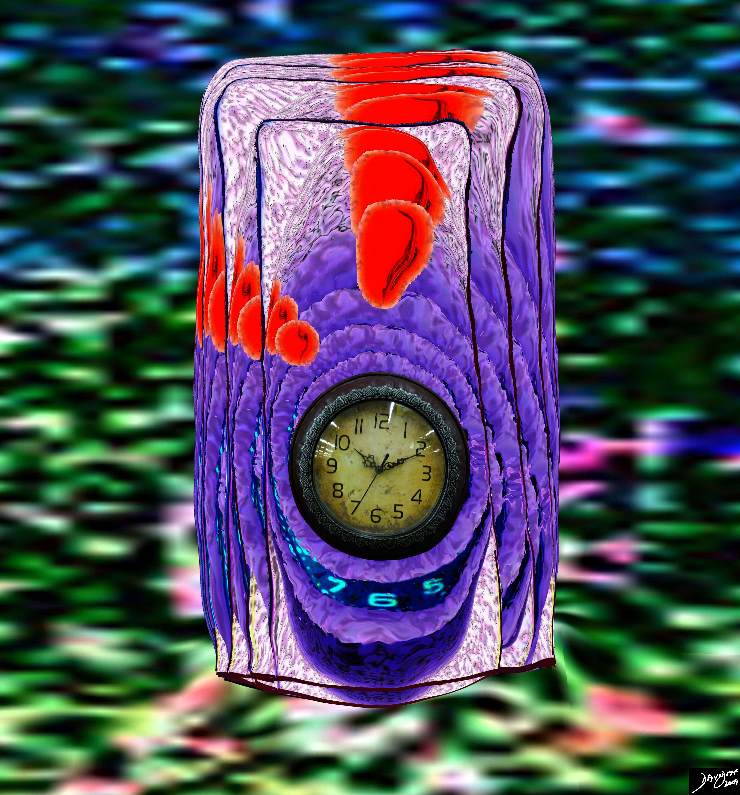
A Single Aberrant Cell – In the Sea of Limited resources |
| 85198j03s.81s The image represents the life of a single set of columnar cells showing a progressioon of generations as the cell lives dies and is regenerated. The orange secretions of the cell are seen in the background of the pink cytoplasm and the purple nucleus. The nucleus of the newesest generation and cell is seen as a clock that has become distorted and time has become disordered. This process is abnormal and is a forerunner of a malignant process. histology time malignancy cancer columnar cell histopathology Davidoff art copyright TheCommonVein.net |
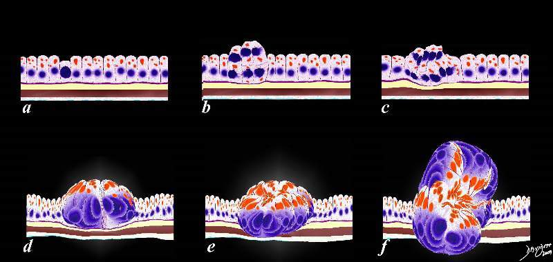
Multiplication of the Rebel Over Time Provides No VAlue for the Community at Large Space Occupation – Pushing and Shoving for Space |
| 85198fc02.8s The image represents the evolution of a single cancer cell (a) that fails to conform to the usual time cycles, grows, multiplies, (b,c,d) and then invades the space of other parts of the tissue like the muscular layer in e and the serosal layer in f. histology time malignancy cancer columnar cell histopathology Davidoff art v |
Rebel in the Community |
| 87160pb06b02b02.81k.8s Different mean space occupation push shove multiplication cancer malignant division bizarre mean loose bonds lack cohesion small hyperchromatic dance movement units to unity individual uniqueness group Davidoff art Davidoff photography copyrightTheCommonVein.net |
The Rebel Creates Progeny by Multiplication |
| 87160pb06b02b02.8k.8s Different mean space occupation push shove multiplication cancer malignant division bizarre mean loose bonds lack cohesion small hyperchromatic dance movement units to unity individual uniqueness group Davidoff art Davidoff photography copyright 2009 all rights reserved TheCommonVein.net |
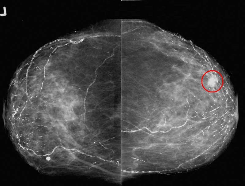
Mammogram Showing a Nodule The Rebel Community |
| 43716r01 hx 82 year old presenting for baseline screening mammogram, positive family history of breast cancer with daughter diagnosed in her 40’s CC views show the spiculated mass in the lateral right breast. breast fx mass fx calcified vascular calcifications ductal calcifications dx lobular carcinoma RX US guided needle loc with surgical excision and XRT breast imaging radiology mammogram mammography Courtesy Priscilla Slanetz MD MPH TheCommonVein.net |
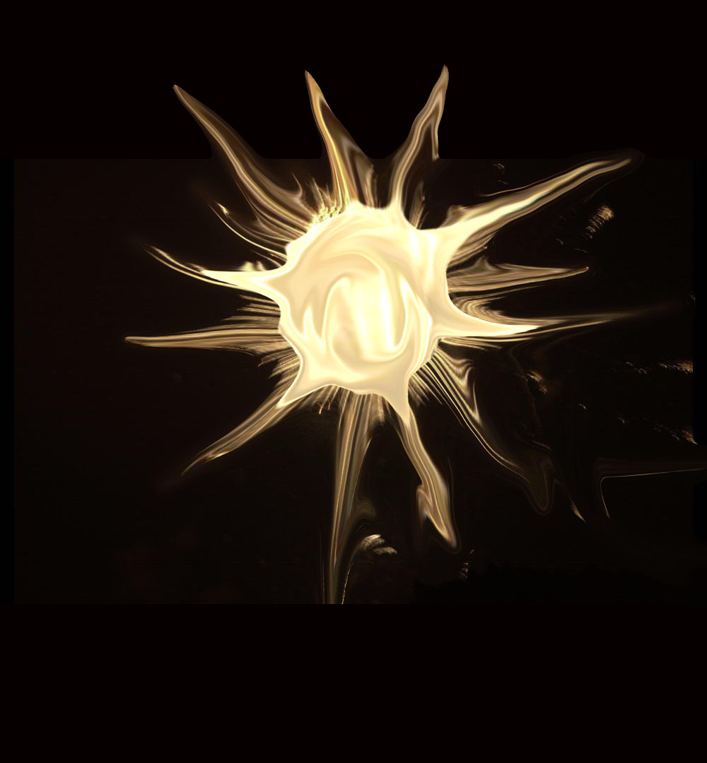
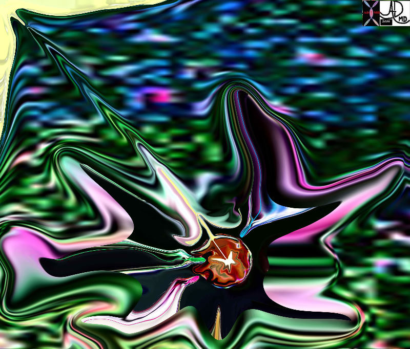
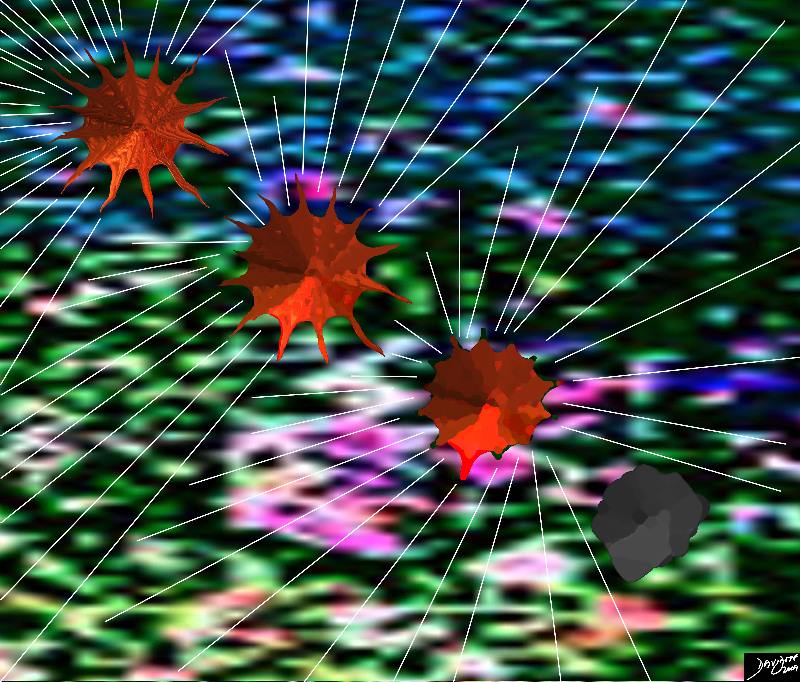
The Spiculated Form of the Enemy |
| 69595b04.800 star stellate corona radiata spiculated aggressive cancer carcinoma Davidoff art TheCommonVein.net |
Breast Cancer |
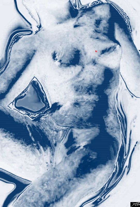
| 78378pb06d.3k.8s breast cancer tiny spiculated lesion too early to palpate visible only to mammogram or MRI Davidoff art TheCommonVein.net |

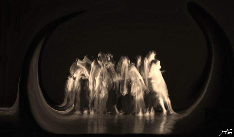
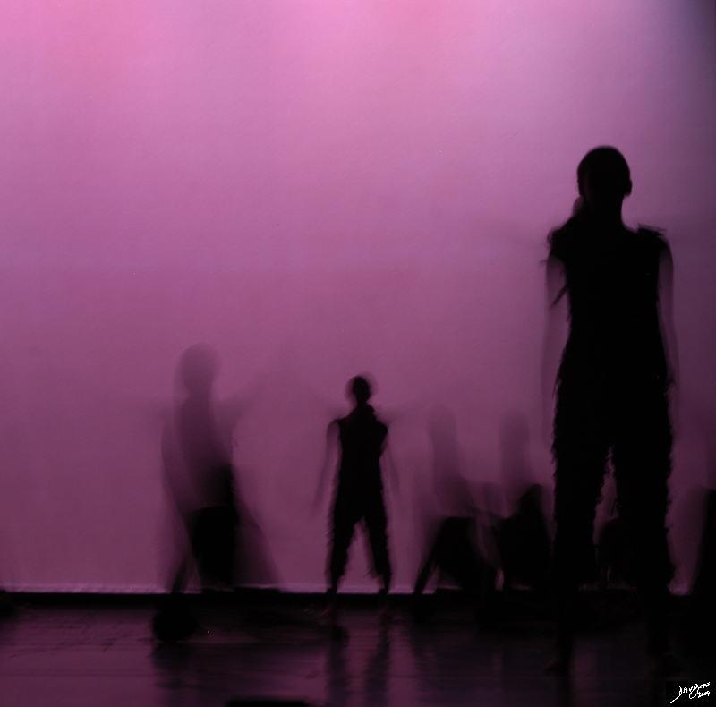
Alone .. So Quietly Desperate… I Cannot Believe it . What About my Children? |
| 87098pb05.8k.8s Wings That Won’t Fly dance movement magic carpet ride units to unity individual uniqueness group Davidoff art Davidoff photography TheCommonVein.net |
Trapped |
| 87119pb08.8k.8s trapped dance movement units to unity individual uniqueness group Davidoff art Davidoff photography copyright TheCommonVein.net |
I told a Friend …We cried |
| 82811pd01.4.8s I need your help Sad news Need your support Bad news Davidoff art Davidoff photography TheCommonVein.net |
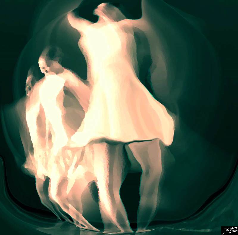
In the Quiet of My Mind – I Searched My Soul |
| 86965pb01.8s faith dance movement arms Davidoff art Davidoff photographyTheCommonVein.net |
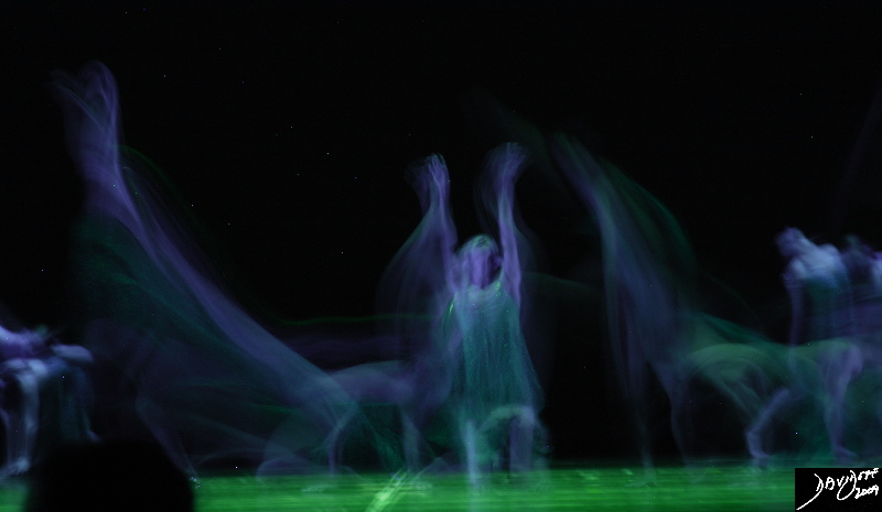
Thinking it Out
Facing it – Finding Out Speaking – Friends Family Doctors Spirtitual healers Others with the Same
Planned Approach for the physical side –
Support, Hope, and Strength |
|
87121pb.8k.8s Davidoff art Davidoff photography copyright TheCommonVein.net |
XRT |
| 87161pb03.8s dance breast cancer XRT radiation therapy Davidoff art TheCommonVein.net |
The Battleground against the Rebel |
| 93002pa.44k.8s cancer treatment chemotherapy web cell death Davidoff art TheCommonVein.net |
Hope |
| 87090pb05.8k.8s dance movement bugs units to unity individual uniqueness group Davidoff art Davidoff photographyTheCommonVein.net |
Hope |
| 87104pb05.8kb.8s breast dance cell to soul units to unity cancer treated cancer free malignant born again freedom joy of life Davidoff art TheCommonVein.net |