Blood Supply
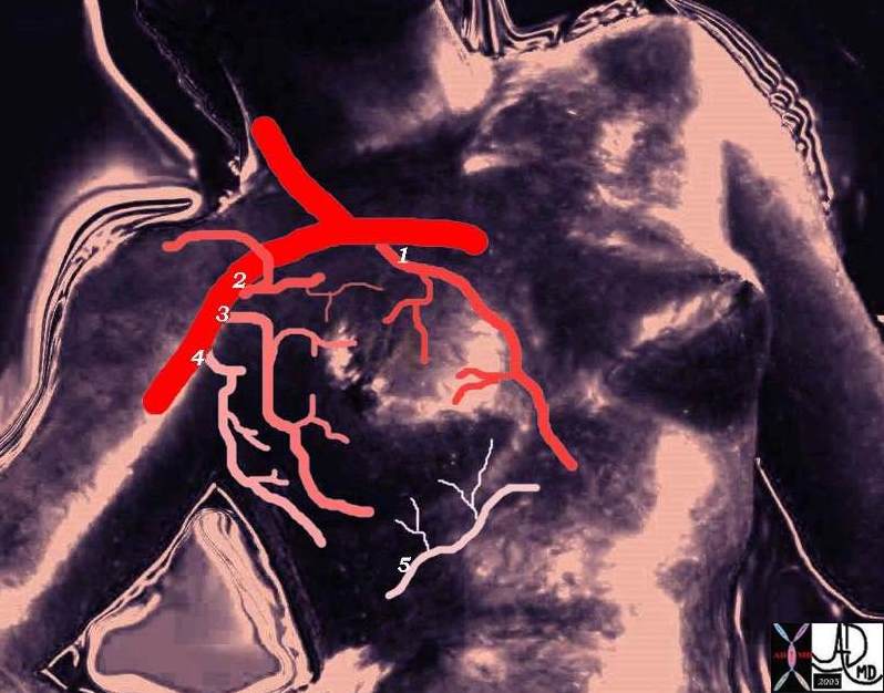
The breast is supplied with blood from 5 sources. The main vessels that supply the breast are the internal mammary artery (internal thoracic) that arises from the subclavian artery and lateral thoracic artery that arises from the axillary artery. The thoracoacromial artery, and the thoracodorsal artery are two other arteries that supply the breast and they both arise from the axillary artery which is a continuation of the subclavian artery. The intercostal arteries of ribs 3-5 are the last of the 5 vessels that supply the breast. They arise posteriorly from the aorta and anteriorly from the internal mammary artery, thus forming a circumferential ring around the chest.
 Blood supply of the breast Blood supply of the breast |
| The blood supply of the breast arises from 5 main sources including the internal mammary artery from the subclavian artery (1), three branches from the axillary artery including the thoracoacromial branch, (2) the lateral thoracic (3) the thoracodorsal artery that arises from the axillary artery and intercostal arteries arising from branches supplying ribs 3-5 (5). Courtesy Ashley Davidoff MD 78378pb09a02 |
The blood of the breast skin depends on the subdermal plexus, which is in communication with underlying deeper vessels supplying the breast parenchyma. There is a particularly rich periareolar plexus. In the world of plastic surgery this rich blood supply allows for a variety of reduction techniques, ensuring the viability of the skin flaps after surgery.

Blood supply of the breast |
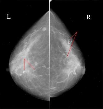
| This mammogram reveals calcified atherosclerotic arteries as depicted by the red markers. Note the course of the vessels is generally from chest wall toward the areola and nipple. Courtesy Priscilla Slanetz MD MPH 42769 |
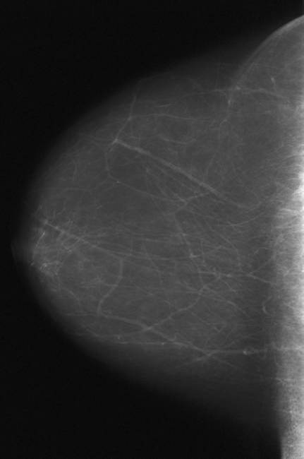
Blood supply of the breastThe uniform vasculature of these fatty breasts can be clearly visualized in this cranio-caudal projection Courtesy Priscilla Slanetz MD MPH 42874 |
 Blood supply of the breast Blood supply of the breast |
| The uniform vasculature of these fatty breasts can be clearly visualized in this cranio-caudal projection Courtesy Priscilla Slanetz MD MPH 42874 |
The Mayo foundation has a 3D arterioscopy movie that takes you on a journey through the arteries of the breast. In my opinion there is not much to be taught by the video but it is fun to feel the technology. arterioscopy 3D movie Courtesy Mayo Foundation After you have taken the ride click the back arrow to return to our module.
Applied Anatomy
Sandrikov and Fisenko showed that Doppler technique was able to demonstrate increased peritumoral vascularity and high velocity in the blood vessels of malignant lesions. McNicholas and colleagues studied blood flow patterns in the internal mammary artery by Doppler technique in patients with benign and malignant disease. They concluded that it was difficult to distinguish the two entities using Doppler technique alone. However when used in combination with the patients age, the size of the lesion and its morphology on ultrasound, distinction was far more accurate. The assessment was also enhanced by the use of mammography. The blood supply to the breast is very rich, and ischemic disease of the breast is almost unheard of. There is however a case report of a patient who developed necrosis of the medial aspect of her left breast following a CABG procedure in which the left internal mammary artery was used as the bypass vessel. (Gonyon)

