As noted the breasts are modified apocrine sweat glands that develop to the same extent in both males and females during embryonic life and are similar structures in both sexes until thelarche when they develop in the female under the influence of the sex hormones to become the organs of lactation providing milk and nourishment for the newborn.
In the female, the breasts continue to change in structure and function throughout life. In the male they have no known function but are still subject to change under the conditions of obesity, and the abnormal influence of estrogen or progesterone. We will focus on the breast of the female for this module.
The breast is made up of radially positioned glands and ducts that converge on the nipple.
There is a wide variation in breast size, breast symmetry, and breast shape. The size of the breasts is usually related to the amount of adipose tissue, but in the younger patient the stromal and glandular tissues may be more dominant. In general adipose tissue makes up 85-90% of the substance of the gland. Together with the connective tissue the adipose tissue surrounds, packs, and supports the glands or lobes.
The shape of the breasts changes during each physiologic phase of the female. In childhood they are flat, while in adolescence they become conical. As the breasts become fully developed the inferior part rounds out, while superiorly they become concave. The discrepancy in shape between the superior and inferior aspects of the breast is probably due to varying lengths and tensions of Cooper’s ligaments – hair sized ligaments that provide support from the skin surface to the chest wall. With the aging process, the ligaments stretch or even break, the breasts lose support and they sag. In 1913 Mary Phelps Jacob a New York socialite, created the brassiere, in an attempt to prevent the dreaded droop. This Western industry capitalized on the social and cultural negative sentiment of the sagging breast. Prior to that, artificial support was in the form of a body corset with whale bones whose creation was attributed in the sixteenth century to Catherine de Medici.
The breasts are the seat of many diseases including fibrocystic change, fibroadenoma, mastitis, and carcinoma. Breast cancer unfortunately is the most common malignancy in women and is second to lung carcinoma as the leading cause of cancer death. In 1960 one in twenty women had the misfortune of developing breast cancer and that figure has increased to one in 8 currently.
Diseases are commonly diagnosed by clinical examination, mammography, ultrasound and biopsy. Clinical concerns are raised mostly when an abnormal mass is felt, or a bloody discharge from the nipple is noted. The continued morphological changes create symptoms that may mimic disease, so that most patients have normal lumpiness in their breasts and many patients have a discharge as normal physiological events. Most small cancers are clinically silent and can only be detected by mammography. Screening mammography starting at age 40 is an essential strategy in the management of breast diseasesther organs, have been a visible structure ever since the birth and evolution of the mammal. Although they have run the gamut of religious, psychological, and erotic environments in history, the study of its internal structure seems to have arrived relatively late. The breast was viewed as sacred (especially pre-1400), erotic (Renaissance and thereafter), political (French Revolution), psychological (as in Freud’s obsession), and medical (including breast cancer and cosmetic surgery).
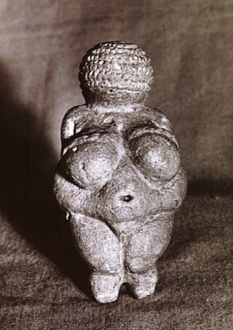
Witcombe a professor of art history at Sweet Briar College in Virginia explored the art and culture of the female figure in ancient civilizations. An electronic version of the work is available (Witcombe). Venus of Willendorf is probably the oldest figurine originating from the stone age (25000BC) The sculpture was found by Josef Szombathy in 1908 in Austria. She is believed to represent a goddess and is characterized by large breasts, abdom

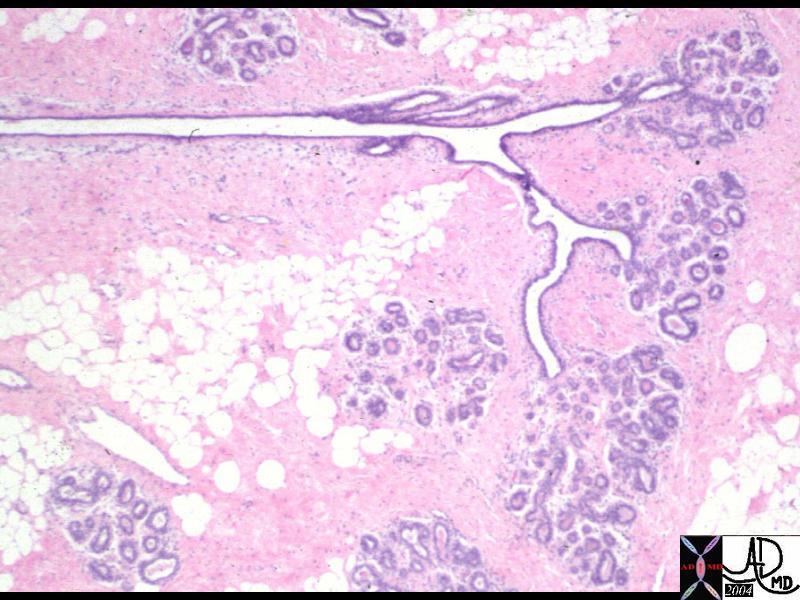
This histological section of the breast reveals part of a lobe subtended by a duct that will terminate in the nipple. There are 15-20 lobes in the breast that are radially distributed around the breast. The structure of the glands of the breast, exemplify a compound branched tubulo-alveolar type gland. (see text for explanation) Courtesy Frank Reale MD Ashley Davidoff TheCommonVein.net 13513

54866


The Goddess of snakes is a well known deity from the Minoan culture of Create from around 1600BCE. (Witcombe image)
54869

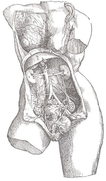
Breast Anatomy Vesalius
Drawing by Vesalius (1514-1564) one of the earliest drawings revealing an interest in the internal structure of the breast From De Corporis Humani Fabrica of 1543g 13497
Classification Based on Structure:
Structurally the breast is part of the skin and can be considered a modified apocrine sweat gland. As discussed above sweat glands are considered apocrine, eccrine or sebaceous in nature. These glands each produce different substances each with different modes of secretion. Apocrine sweat glands produce a thick secretion which is secreted into the duct by pinching off the secretion from the apex of the gland, while eccrine glands secrete a watery fluid (sweat) by an active mechanism directly into the duct by the classical secretory mechanism without losing part of its cytoplasm in the process. The sebaceous gland produces sebum and is associated with hair follicles. The sebaceous gland and the apocrine gland only become functional during adolescence as the individual matures sexually. The apocrine glands are associated with the hair follicles of the axilla, pubic area, and breast and produce a secretion that contains pheromones. The sebaceous glands are associated with hair follicles elsewhere in the body. The areolar glands of Montgomery are sebaceous glands that enlarge during pregnancy secreting an oily sebaceous fluid to act as a protective lubricant for the areola and the nipple
Classification Based on Function:
The breast is an exocrine gland meaning that its secretion is transported into a ductal system. An endocrine gland on the other hand (for example the adrenal gland) secretes its products into the blood stream. The pancreas for example has both endocrine components (insulin secreted into the blood) and exocrine components (digestive enzymes secreted into the ducts).
Functionally the breast is part of the reproductive system. Its primary role is to provide nutrition for the newborn infant, but it also provides essential cognitive and emotional aspects for the infant.
The breast acts as a secondary sex organ meaning that it is an organ that is not directly related to reproduction but enables physical distinction between the sexes. Voice depth and facial hair are other secondary sexual characteristics.
Classification Based on Histology:
The breast is part of the skin and its glands. The skin contains two major layers – the epidermis, and dermis. The epidermis contains the hair follicles, sweat glands, sebaceous glands, nails and the mammary glands, while the dermis below consists of dense connective tissue.
The functioning component of a gland is the epithelium which in the case of an exocrine gland is organized around a ductal system. The epithelium in the breast is cuboidal meaning that the cells are almost square in shape. The shape however will vary from being flat to columnar depending on the stage of functional stimulation. Under the cuboidal epithelium in the lobules is a second layer of myoepithelial cells.
As a gland that has a lobular configuration it is considered to be a compound tubuloalveoalar gland. As a “compound” gland it implies that the ductal portion is branched. As a “tubuloalveolar” gland it is implied that the secretory element of the gland has both tubular and alveolar (grape-like) elements.
Classification Based on Embryology:
We have learned above that the breast is part of the skin which is made up of the superficial epidermis and the deeper dermis. The epidermis is derived from the ectodermal germ layer and the dermis arises from the
General
The female breast is a dynamic structure that from month to month, with visible and palpable structural changes. These changes become more dramatic during pregnancy and lactation.
The structural changes that are visible and palpable to the patient are often combined with the ever present fear of malignant disease.
Many patients therefore have multiple visits to their clinicians and radiologists, to allay or confirm their fears. An improved knowledge of the anatomy and the associated structural changes that occur with the physiology would serve all well.
